Research
The ability to invade host tissues and metastasize is the major cause of cancer-related death. During tumor invasion, metastasizing cells disrupt normal cell-cell and cell-matrix contacts and acquire a migratory, invasive phenotype. Thus modulation of cell-cell and cell-matrix adhesive events likely plays a critical role in tissue remodeling during tumor progression.
Subsequent alterations in cellular architecture mediated by modified extracellular matrix (ECM) attachments induce expression of proteinases that degrade ECM proteins, facilitating migration through the modified tissue to establish metastatic foci and removing matrix constraints that normally limit proliferation.
Although malignant cells produce a spectrum of matrix-degrading enzymes, predominant among these are extracellular serine and metallo-proteinases. Current research centers on regulation of these proteinase families in two model systems: epithelial ovarian carcinoma and squamous cell carcinoma of the oral cavity. Ongoing research utilizes an integrative approach involving examination of 2-dimensional (2D) and 3D tissue culture systems and organotypic cultures complemented by murine xenograft models and analyses of human tumors.
Understanding the molecular mechanisms by which tumor cells orchestrate multiple micro environmental cues to regulate metastasis is the major focus of the Stack Lab.
View research publications here
Epithelial Ovarian Carcinoma
Epithelial ovarian carcinoma is the leading cause of death from gynecologic malignancy, as 75% of women with this disease succumb to complications resulting from disseminated intra-peritoneal metastasis. Thus, strategies aimed at the prevention of metastasis would generate immediate clinical impact.
The initiating events in the development of ovarian carcinoma are poorly understood. However, ovarian carcinoma has a unique metastatic mechanism requiring reversible modulation of cell-cell and cell-ECM contacts that involves shedding of cells from the primary tumor followed by subsequent intra-peritoneal adhesion, invasion, and proliferation. Unlike most highly metastatic tumors, the majority of women with advanced intraperitoneal disease have no clinically apparent lymphatic or hematogenous metastases, implying that a novel mechanism for metastasis is operative in ovarian cancer.
Further, as metastases are largely confined to the peritoneal cavity, microenvironmental factors that modulate intraperitoneal adhesion, motility, and invasion play a predominant role in ovarian pathobiology. Current studies are investigating the role of extracellular proteolysis in ovarian cancer early dissemination and intra-peritoneal anchoring, adhesion-modulated signal transduction and alteration of gene expression, and the role of microenvironmental factors including peritoneal adiposity and ascites fluid in progression to metastasis.
Oral Squamous Cell Carcinoma
Oral squamous cell carcinoma (OSCC) is the most common malignancy of the oral cavity, causing more deaths than any other oral disease. The 5-year survival rate has not improved appreciably in over 20 years, remaining at a low 50%. These statistics reflect our limited understanding of the molecular events that govern disease progression.
Treatment of advanced OSCC is associated with high mortality, resulting from local, regional, and distant metastasis; however, the cellular and biochemical factors that underlie OSCC dissemination are poorly understood. In the absence of reliable molecular markers for use in early detection or as prognostic indicators, treatment failures remain difficult to predict. Thus, a more detailed analysis of the molecular events that contribute to OSCC metastasis are a necessary prerequisite to the development of novel early detection and treatment strategies that favorably impact the survival of OSCC patients.
To this end, our recent studies have utilized cDNA microarray and microRNA array analyses of aggressive OSCC to identify changes associated with OSCC primary tumors and lymph node metastases. These studies are focused on identification of prognostic biomarkers and understanding the molecular events in OSCC progression. An additional research effort is in the area of HPV-associated oropharyngeal cancer (OPSCC). A marked >200% increase in OPSCC has been noted in the last 15 years in a younger age cohort (20-40 year olds). Current efforts are focused on microRNA profiling of OPSCC, understanding the link between microRNA expression, proteomic profiles, and disease progression; and development of microfluidics-based early detection technologies.
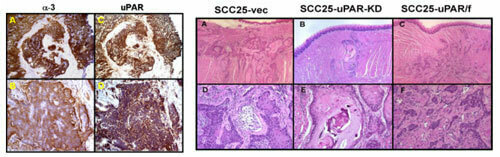
Oral squamous cell carcinoma can occur in multiple sites in the oral cavity and oropharynx. The center and right panels above show the murine orthotopic xenograft model of aggressive OSCC. These present as infiltrative lesions with ill-defined borders with clusters of hyperchromatic cells with a high nuclear to cytoplasm ratio. There is cytologic atypia and mitotic figures are easily identified. In vivo imaging of fluorescently-tagged OSCC cells can be used for longitudinal imaging of tumor progression.
Investigators in the Harper Cancer Research Institute are dedicated to conducting innovative and integrative research that confronts the complex challenges of cancer.